Description
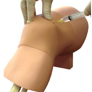
Skill Development
- Learn to ballot or milk the suprapatellar pouch
- Needle aspiration and injection techniques
- Palpating anatomic landmarks significant to the procedure
- Realistic tactile feedback throughout procedure
Features
- Ultrasound compatible
- Highly durable replaceable tissue for multiple uses
- Realistic tactile feedback includes:
- Sensation of bony contact if needle hits the patella
- Sensation of bony contact if needle hits the femur
- Inability to aspirate the syringe while the needle tip is in the soft tissue superficial to the join capsule
- Easy aspiration of joint fluid into the syringe once entry has been achieved
- Fluid can be left clear or be colored if desired
- Ability to increase or decrease size of effusion with up to 60 cc of fluid
Components
- Arthrocentesis Base, Replaceable Tissue (ARCT-20), 60ml luer-lock syringe, Instruction Sheet
Dimensions
- Size: 13″ h; 8.5″ w; 7″ d
- Weight: 7.5 lbs


