Ultrasound Simulation Manikin
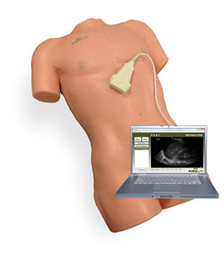
SNM-20; SonoMan Training System: (PC not included)
This ultrasound trainer provides an affordable platform for teaching students how to read diagnostic ultrasound imaging. The system includes a soft tissue body form with internal and external landmarks, and a simulated prob. The torso has 258 unique probe locations providing normal and abnormal images in each window. The system’s software platform allows users to expand SonMans’s capabilities with a variety of training modules. Each of the exam modules contain two normal patients and three to five abnormal pathologies.
System Modules
- FAST Module
- AAA Module
- ECHO Module
- RENAL Module
- GALLBLADDER Module
- eFAST Module
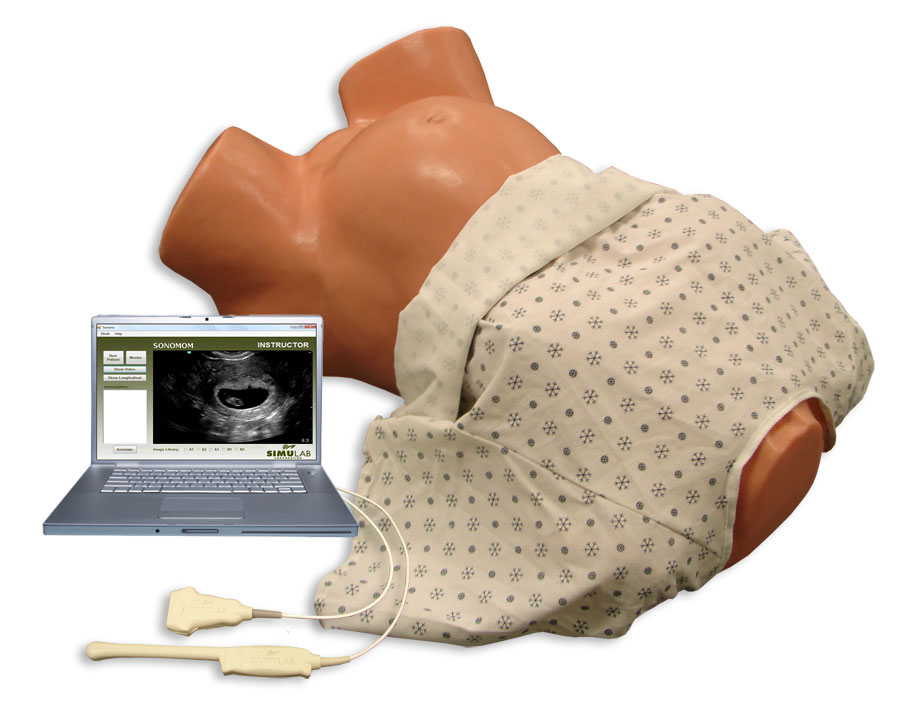
SMM-10; SonoMom Training System: (PC not included)
This ultrasound trainer provides an affordable platform for teaching students how to read obstetric ultrasound imaging for three different exams. SonoMom’s soft tissue body form includes internal and external landmarks, and two simulated probes for abdominal and endocavity scans. The torso simulates a pregnant patient from 6-weeks to 40-weeks gestational age, and has 147 unique probe locations. The 1st Trimester Exam Module includes; two non-pregnant normal patients and 13 pregnant patients with normal and abnormal findings.
System Modules
- 1st Trimester
- To be released
- 2nd Trimester
- 3rd Trimester
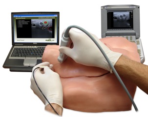
ISBS-10: Nerve Block Training System
INTERSCALENE / SUPRACLAVICULAR BLOCK TRAINER
The Interscalene / Supraclavicular Block Trainer ultrasound compatible and has built-in nerve stimulation simulation. It incorporates SimuLab’s SartTissue(patent pending), which allows the tissue to communicate with a PC, showing the user when the needle or cathetyer are proximal to or in contact with C5, C6, or C7 nerve.
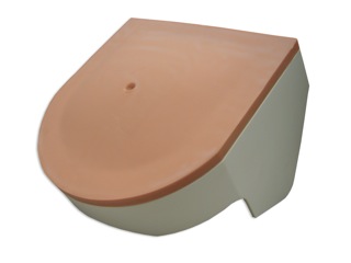
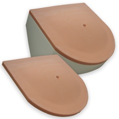
FBT-10; Foreign Body Ultrasound Trainer
The Foreign Body Ultrasound Trainer contains various foreign bodies at varying depths allowing users to practice using ultrasound to identify and measure items, which include a Nail, a Needle, a Glass shard, a Bullet, and a Metal fragment.
The Foreign Body Instructor Model provides a key for the Foreign Body Ultrasound Trainer, revealing where the various foreign bodies are located within the trainer for further use in instruction
LPE-10; Lumbar Puncture / Epidural Trainer:
The Lumbar Puncture / Epidural Trainer is an ultrasound compatible trainer that includes the lumbar vertebrae, iliac crest, ligamentum flavum, the epidural space and dura. The trainer can be positioned in an upright sitting or a lateral decubitus position. The lumbar puncture procedure allows the user to penetrate the subarachnoid space and CSF is present when the stylet is removed. the epidural procedure allows the user to enter the epidural space and insert an epidural catheter.
Available with four different replaceable tissues
- Normal Adult Tissue
- Obese Adult Tissue
- Geriatric Adult Tissue
- Geriatric Obese Tissue
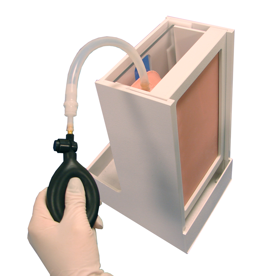
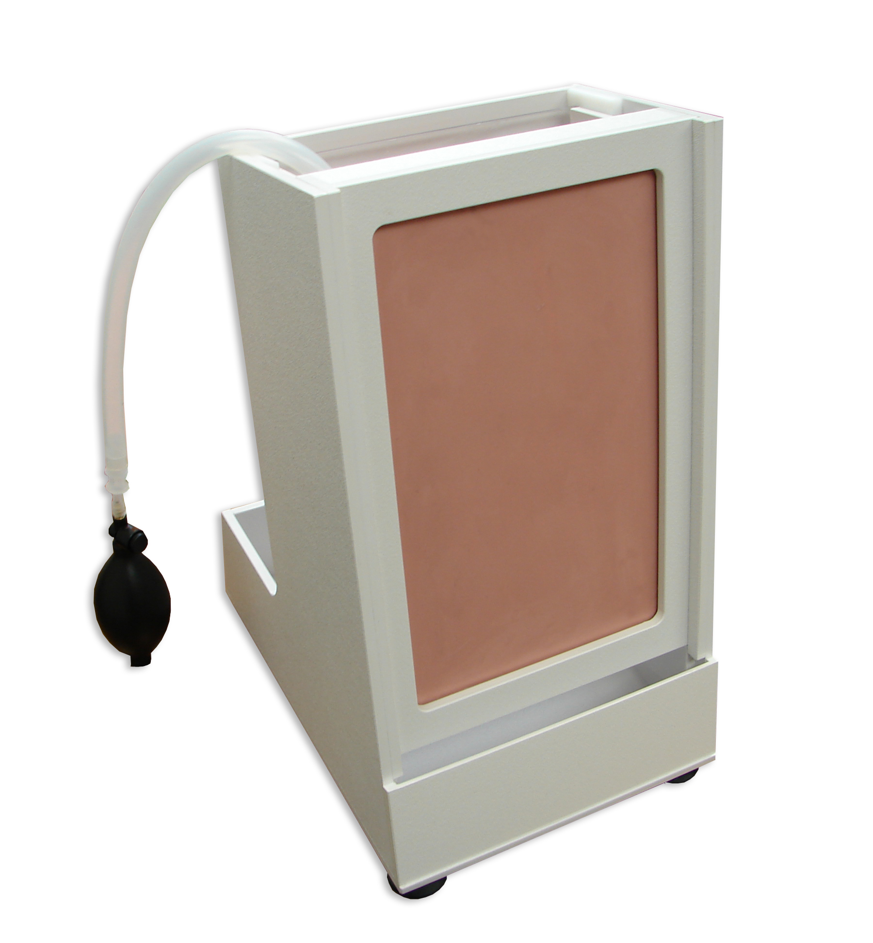
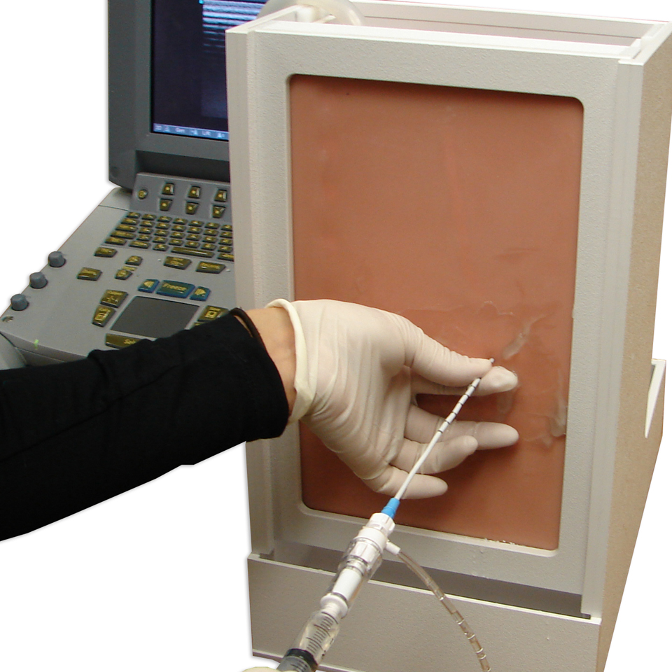
THM-20; Ultrasound Thoracentesis Model:
The Ultrasound Thoracentesis Model simulates a partial torso with anatomical landmarks incuding the scapula, rib, diaphragm, pleural cavity, and lung. The simulated lung can seen as an echogenic structure with an inflation mechanism to adjust the size of the pleural effusion. The open top allows the instructor to provide feedback on procedural concepts by allowing students to visualize the catheter depth and placement when inserted into the pleural cavity. A positive fluid flow then offers users feedback when pleural effusions are accurately accessed. The trainer is designed to be filled with fluid and is self-sealing for multiple procedures.
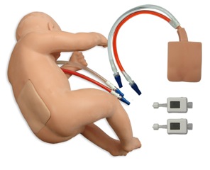
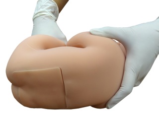
LPB-10: LumbarPunctureBaby Training System
The infant lumbar puncture trainer simulates a two week old infant that can be positioned either lateral or decubitus. The flexible body form is anatomically correct with a partial iliac crest and umbilicus. The replaceable tissue has an articulating L3 – L5 vertebrae with a partial sacrum and the gluteal crest. Each tissue includes a spinal cord filled with simulated CSF and the epidural venous plexus filled with simulated blood providing the user with both a positive or negative response.
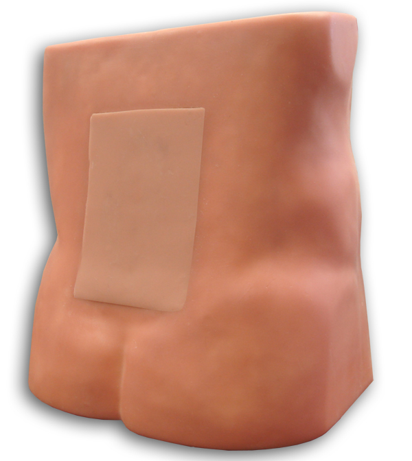
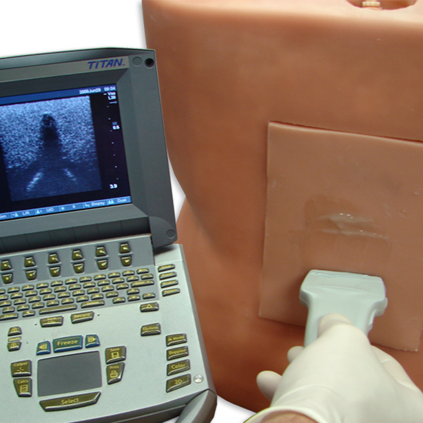
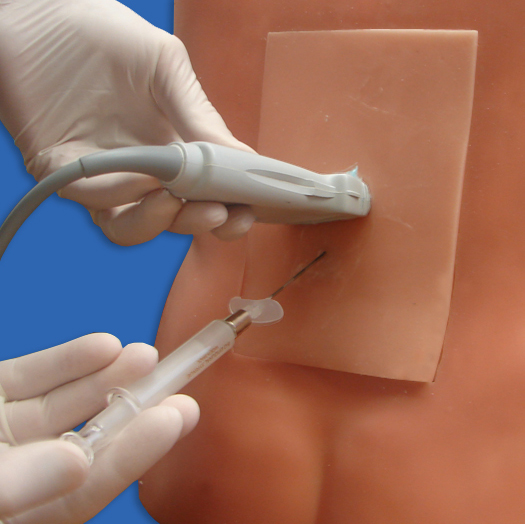
PAC-10: Ultrasound Paracentesis Model
This ultrasound compatible model allows for procedural accuracy when performing a paracentesis. The anatomically correct model features a distended Abdomen and allows the procedure to be performed with or without ultrasound and on either of the recommended areas: the midline below the umbilicus or medial 4 to 5 cm above the anterior superior iliac spine. The model’s anatomy includes superficial epigastric vessels, partial liver and partial spleen, rectus abdominal muscles, and mesentery intestines and allows for removal of up to one litre of fluid from the peritoneal cavity in the abdomen.
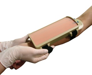
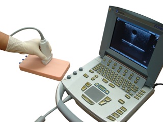
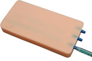
VPP-80: Venipuncture Pad
The Venipuncture Pad, which is ultrasound compatible, are perfect for practicing venipuncture or peripheral catheterization on three varying sized veins filled with blue fluid. The three veins in the pads are slightly visible through the skin surface on one side and obscured by thicker tissue on the other side.
VTH-10: Venipuncture Tissue Holder